
A sharp pain on the edge of the wrist, on the thumb side. It flares up when you lift your child, when you twist open a jar, when you pour from a pan. Sometimes it radiates into the forearm.
De Quervain's tenosynovitis is very common and responds well to treatment. But it has a feature few articles mention: it sometimes fails to respond even to well-conducted treatment, and that failure has a precise anatomical explanation. This is the thread running through this article.
What is really happening in your wrist
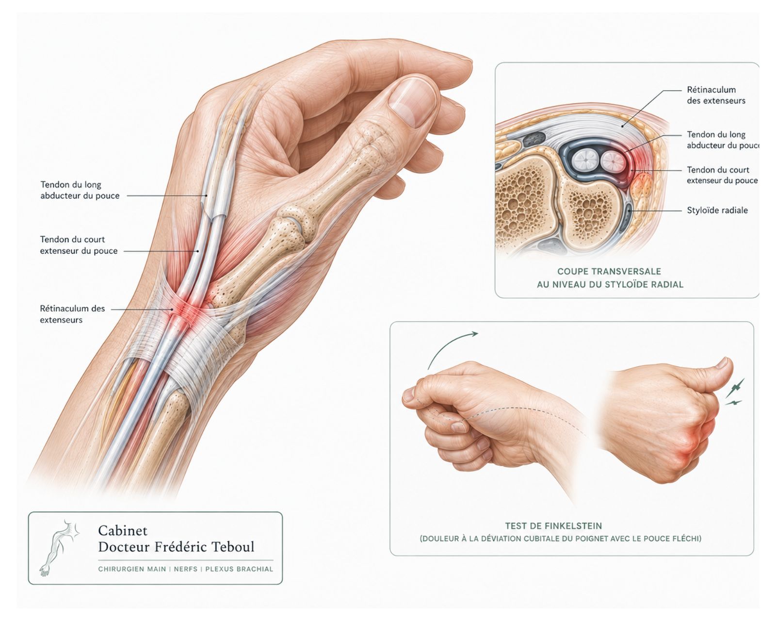
On the outer edge of the wrist, just above the bony prominence you can feel at the base of the thumb — the radial styloid — two tendons pass through a narrow canal called the first dorsal compartment.
These two tendons are the abductor pollicis longus and the extensor pollicis brevis. They are what spread the thumb outward and pull it backward. The canal that holds them is lined by a fibrous sheath pressed against the bone.
In De Quervain's tenosynovitis, this canal narrows and the sheath thickens. The tendons no longer glide freely: every thumb movement becomes friction, and the friction becomes pain.
It is not exactly tendinitis
The common term is « wrist tendinitis ». It is misleading, for the same reason as in trigger finger: samples taken during surgery show not so much acute inflammation as a degenerative reshaping of the sheath, with disorganized collagen and a thickened wall.
This detail has a practical consequence: it explains why oral anti-inflammatory drugs are often disappointing, and why treatments that act on the mechanics — a splint, adapting how you use your hand, opening the canal — give better results. See the trigger finger article.
The partition: what explains most treatment failures
This is the most important point in this article, and it is almost always missing from pages about this condition.
In a large proportion of people affected, the first compartment is not a single canal: a vertical partition — a septum — divides it into two separate tunnels, one for each tendon.
Why this is decisive
If an injection is given without knowing there is a partition, the medication spreads into only one of the two tunnels. The other stays completely unchanged — and the pain persists. The treatment did not fail: it simply did not reach the right target.
The same reasoning applies to surgery: an opening that left one sub-compartment closed would not solve the problem.
This partition cannot be guessed from the clinical examination. It shows up on ultrasound. That is what makes this exam far more than a simple confirmation of the diagnosis in this condition.
Who is affected
De Quervain's tenosynovitis mainly affects middle-aged adults, with a clear predominance in women.
The situations that come up most often:
The postpartum period
This is a category of its own: lifting an infant under the arms, thumbs spread wide, dozens of times a day, stresses exactly the tendons involved. Hormonal changes and fluid retention are also thought to contribute.
Repetitive movements
Movements that combine bending the wrist and spreading the thumb: gardening, racket sports, golf, DIY work, heavy smartphone use.
Inflammatory diseases
In particular rheumatoid arthritis.
A direct blow
To the outer edge of the wrist.
If you have just given birth. This form is common and has a good prognosis, but it raises a specific question: which treatments are compatible with breastfeeding. It deserves to be addressed openly during the consultation rather than simply endured.
Symptoms, and what not to confuse them with
The pain sits on the outer edge of the wrist, just above the radial styloid. It is sharp, sometimes stabbing, and triggered by very recognizable movements: gripping, lifting, twisting open a jar, wringing out laundry, carrying a child.
It can radiate into the thumb or travel up the forearm. A small tender swelling can sometimes be felt above the styloid. At first the pain appears only with effort; over time it can become nearly constant.
Four neighboring pains that should not be confused
Thumb-base osteoarthritis causes pain lower down, at the base of the thumb itself, not above the styloid. It is also triggered by opening a jar — hence the frequent confusion. See the thumb-base osteoarthritis article.
Carpal tunnel syndrome causes nighttime tingling in the fingers, not mechanical pain on the edge of the wrist. See the carpal tunnel article.
A bone problem or a cyst can also cause pain on the radial side of the wrist.
Intersection syndrome, which is rare.
These distinctions are not a matter for self-diagnosis: they call for an examination by a hand surgeon, who assesses the hand and wrist as a whole.
Diagnosis — and a test almost everyone describes backwards
The diagnosis is clinical. The examination finds pain specifically on pressing over the first compartment, above the radial styloid, and sometimes a noticeable thickening of the sheath.
Two maneuvers are used, and they are very widely confused — including in teaching textbooks.
The confusion is long-standing and well documented. A study of 36 people without symptoms — 72 wrists — compared the two maneuvers: the Finkelstein test proved more accurate, with better specificity, significantly fewer false positives, and less discomfort. The authors recommend keeping it as the reference clinical test. Finkelstein vs Eichhoff
Why this distinction matters to you. An Eichhoff test can be painful in someone who does not have De Quervain's. A diagnosis based on this maneuver alone therefore risks treating a condition you do not have — or missing the real cause of the pain.
The role of ultrasound
It shows the thickening of the tendons and sheath, any fluid buildup, and above all the presence of a partition and distinct sub-compartments. It also allows an injection to be guided precisely.
MRI is almost never needed; it is reserved for atypical cases or for looking for another cause. X-rays are usually normal.
Treatments: what the comparisons really show
This is where the information available online is the vaguest — and also where the recent data are the most interesting.
A network meta-analysis published in 2024 compared all the non-surgical options against one another, drawing on 14 randomized trials, over three time frames: six weeks, six months, and one year. Level I evidence. 2024 meta-analysis
The authors' conclusion, not to be distorted
Corticosteroid injection combined with short-term immobilization remains the main, effective treatment for De Quervain's tenosynovitis. Shockwave therapy can be considered a second-line option. Injections on their own should be approached with caution, given the lack of any substantial benefit demonstrated over placebo.
In other words: it is not the injection that is at fault, it is the injection on its own. Combined with immobilization, it remains the standard of care.
The point that changes everything: where you inject
A second network meta-analysis, published in 2023, supplies the missing piece.
Among all the options compared for short-term pain, the one with the highest probability of being the most effective was an ultrasound-guided injection targeted only at the compartment of the extensor pollicis brevis, when sub-compartmentalization is present. 2023 meta-analysis
Conversely, a placebo injection had the highest probability of being the least effective, followed by wearing a splint.
The two meta-analyses echo each other. An injection on its own disappoints in the short term — but an ultrasound-guided injection aimed at the right sub-compartment comes out on top. It is not the medication that makes the difference: it is knowing where to place it.
And shockwave therapy?
It ranks first in the short and medium term in the 2024 meta-analysis, which is a notable result. The authors, however, position it as a second-line option, not a first-line treatment. Its exact place in a care pathway still needs to be defined case by case.
First-line measures
They remain entirely worthwhile, provided they are understood for what they are: supportive measures rather than stand-alone cures.
Adapting how you use your hand — changing the way you lift a child, open a jar, hold a phone.
A splint immobilizing the thumb and wrist, especially at night and during activities that trigger the pain. On its own, it was no better than placebo in the short term in the comparisons; combined with an injection, it is part of the standard treatment.
Anti-inflammatory drugs, for a short period.
Rehabilitation: tendon-gliding exercises, gentle stretches, gradual strengthening.
Surgery
It is considered when conservative measures and one or two injections have not been enough, or when the pain is very disabling.
The procedure consists of opening the compartment to free the passage of the tendons. Two technical requirements determine the outcome:
Identifying and protecting the sensory branch of the radial nerve, which runs just above the compartment. Injuring it is the complication specific to this procedure.
Checking that no sub-compartment is left closed. This is the point made at the start of this article: an incomplete opening leaves a tendon compressed and the pain persists.
Published series report high cure and satisfaction rates. The complications described are irritation or a neuroma of the sensory branch of the radial nerve, tendon instability if the opening is too wide, and, more rarely, prolonged residual pain.
After the procedure
The procedure is performed under regional anesthesia as an outpatient, through a transverse (cosmetic) incision. Except in particular cases there is no post-operative immobilization, with immediate thumb movement as pain allows. Rehabilitation may be needed in patients who are apprehensive about moving the thumb after a long-standing case of De Quervain's tenosynovitis.
In pregnant patients and while breastfeeding, treatment relies mainly on immobilization because corticosteroid injections are contraindicated; surgery may be considered in especially painful cases.
The special case of the postpartum period
This form deserves a separate answer. A new mother faces three constraints at once: a triggering activity she cannot avoid — carrying her child —, a possible question of whether treatments are compatible with breastfeeding, and limited availability for follow-up.
The good news is that this form has a good prognosis. But the management cannot be a simple « rest your wrist ».
Frequently asked questions
Glossary
The narrow canal on the outer edge of the wrist in which the two tendons involved glide.
The bony prominence of the wrist, on the thumb side, the landmark for the pain.
An anatomical partition dividing the canal into two separate tunnels. Its presence explains many treatment failures.
A maneuver in which the examiner grasps the thumb and draws it toward the little finger. Not to be confused with the Eichhoff test.
A treatment delivering mechanical pulses through the skin, without injection or incision.
References
Advancements in de Quervain Tenosynovitis Management: A Comprehensive Network Meta-Analysis. J Hand Surg Am. 2024. PMID 38613563 — 14 randomized trials, Level I evidence
Challoumas D, Ramasubbu R, Rooney E, Seymour-Jackson E, Putti A, Millar NL. Management of de Quervain Tenosynovitis: A Systematic Review and Network Meta-Analysis. JAMA Netw Open. 2023;6(10):e2337001. PMID 37889490
Finkelstein H. Stenosing tendovaginitis at the radial styloid process. J Bone Joint Surg. 1930;12:509-540
Leao L. De Quervain's disease: a clinical and anatomical study. J Bone Joint Surg Am. 1958;40-A(5):1063-1070 — documented origin of the confusion between the two tests
Comparative study of Finkelstein / Eichhoff in 36 asymptomatic subjects (72 wrists) — the Finkelstein test proves more specific, with fewer false positives
Goubau JF, Goubau L, Van Tongel A, Van Hoonacker P, Kerckhove D, Berghs B. The wrist hyperflexion and abduction of the thumb (WHAT) test. J Hand Surg Eur Vol. 2014;39(3):286-292
Huisstede BMA, Coert JH, Fridén J, Hoogvliet P; European HANDGUIDE Group — European consensus on the management of De Quervain's tenosynovitis
Prevalence of the intra-compartmental septum
Success rates of injections and surgery
Histological study of the degenerative nature
Content provided, reviewed, and approved by Dr Frédéric Teboul, hand, brachial plexus, and peripheral nerve surgeon, member of the Académie Nationale de Chirurgie.
Last updated: 20 August 2026
Disclaimer. This article is for information only. It does not replace a medical consultation. Only a clinical examination can establish a diagnosis and choose a treatment suited to your situation.
Author of this publication
Every piece of content is signed by its author and reviewed before publication.
 Author
Dr Frédéric Teboul
Hand surgeon — specialist in the brachial plexus and peripheral nerves
Author
Dr Frédéric Teboul
Hand surgeon — specialist in the brachial plexus and peripheral nerves