
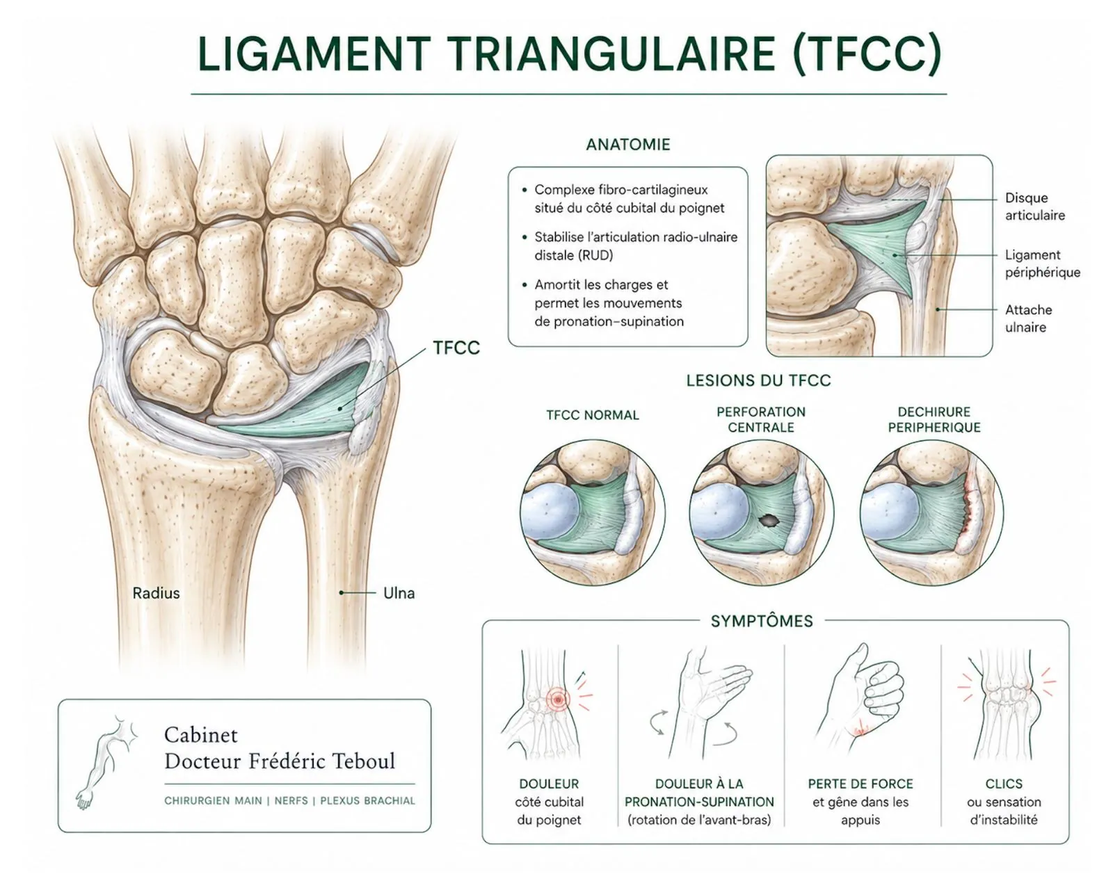
Pain on the little-finger side, at the edge of the wrist. It flares up when you turn a key or a door handle, when you lean on your hand, or when you strain in rotation. Sometimes a click, a feeling of instability.
This inner-wrist pain is common and often mislabeled. In a large share of cases, it comes from the triangular fibrocartilage. This article explains what it is, how the diagnosis is made, and why two simple questions determine the entire treatment — far more than the exact type of injury.
The triangular fibrocartilage, or TFCC: the shock absorber of the inner wrist
On the inner side of the wrist, on the little-finger side, the forearm ends in two bones: the radius, on the thumb side, and the ulna, on the little-finger side. Between the end of the ulna and the wrist bones lies a complex fibrocartilaginous structure, the triangular fibrocartilage — in English, triangular fibrocartilage complex, which gives the abbreviation TFCC that you will see everywhere.
It is not a single ligament but a complex: a central disc, similar to a small meniscus, and peripheral ligaments that anchor it to the bone. It performs two distinct functions, and this distinction is the key to the whole article.
Function 1 — cushioning
The central part, the disc, absorbs and distributes the loads that pass through the inner wrist. It is a cushion.
Function 2 — stabilizing
The peripheral part, especially at its deep anchor on the ulna — the area known as the fovea — holds the two forearm bones together and provides the stability of their joint, particularly during rotational movements.
Remember this duality: depending on whether the injury affects the cushion or the stabilizing anchor, the problem and the treatment are not the same.
Two very different origins
Triangular fibrocartilage injuries fall into two broad families, which it is essential to distinguish because they do not call for the same response. Palmer 1989
Traumatic injuries
They occur during a specific injury: a fall onto the rotating hand, a forced twisting movement of the wrist, sometimes in the wake of a wrist fracture. They tend to affect young, active people, and this is where the question of stability arises.
Degenerative injuries
They result from gradual wear of the disc, with no single injury. They most often arise in a particular setting: an ulna that is slightly too long relative to the radius, which repeatedly bumps against the wrist bones and wears the ligament from within.
This is called positive ulnar variance : it is the finding that shifts the strategy, as we will see below.
Symptoms
The hallmark symptom is pain on the inner side of the wrist, on the little-finger side, triggered or worsened by:
Rotational movements of the forearm — turning a key, a handle, a screwdriver, unscrewing a cap.
Leaning on the hand, palm down, with the wrist bearing weight — getting up from a chair, doing a push-up.
Bending the wrist toward the little finger, especially under load.
Sometimes there is also a click or a catching sensation during rotation, a loss of grip strength, and a feeling of instability — the sense that "something is moving" at the edge of the wrist. This last sign is important: it points to involvement of the stabilizing part.
Diagnosis
The clinical examination seeks to reproduce the pain through specific maneuvers and, above all, to answer the first of the two decisive questions: is the wrist stable?
One maneuver in particular — checking for abnormal movement of the end of the ulna by pinching it between two fingers — tests this stability. An abnormally mobile ulnar end signals involvement of the deep anchor, the one at the fovea. This is something imaging alone does not always show, which is why the examination matters.
Imaging
It does not show the ligament, but it answers the second decisive question: is the ulna too long? It measures ulnar variance, often on an image taken in a specific position.
Sometimes with an intra-articular injection of contrast (MR arthrography), it shows the ligament and pinpoints the tear.
Examining the wrist with a miniature camera remains the most reliable test: it sees the injury directly, tests the strength of the anchor, and allows treatment in the same session.
The two questions that drive the treatment
You often read complex classifications, with types and subtypes. They help the surgeon describe an injury precisely, but to understand your situation, everything comes down to two questions.
These two questions are independent: a wrist can be unstable without a long ulna, have a long ulna without instability, show both, or neither. It is their combination that shapes the treatment — not the technical name of the injury.
Treatment, question by question
First, without surgery
Many injuries, especially recent ones without instability, improve without surgery. Initial management most often combines:
Rest and adapting your movements, avoiding forced rotation and painful weight-bearing.
Immobilization with a splint or brace, to rest the wrist and forearm rotation.
Anti-inflammatory medication, for a limited time.
A corticosteroid injection into the joint, which can provide relief.
Rehabilitation (physical therapy), once the pain has settled, to restore strength and control of the wrist.
This non-surgical treatment is often tried first, particularly when the wrist is stable. Its duration and role are discussed case by case.
If the disc is worn but the wrist is stable: clean it out
For a central tear, in the cushion, without instability, the standard procedure is arthroscopic debridement: the tear is trimmed through the camera, removing the damaged part that catches, without repair — because this central area, which has a poor blood supply, heals poorly anyway.
A note of honesty that few websites give
Debridement gives good relief and allows a quick return to activity — in one review, about nine out of ten patients went back to work. But it does not make everyone pain-free: in the same review, a little under half were completely free of pain. It is a good procedure, not a guarantee.
And above all: if the ulna is too long, cleaning out alone often fails — in up to a quarter to a third of cases — because the cause has not been treated. The ulna then has to be shortened.
If the wrist is unstable: repair the anchor
When it is the deep anchor, on the fovea, that is torn — that is, when the wrist is unstable — cleaning out is not enough: the ligament has to be reattached to the bone. This peripheral area, by contrast, has a good blood supply and heals well.
The repair can be done arthroscopically or through open surgery; studies show no clear advantage of one approach over the other in terms of pain, function, and mobility. A figure to set expectations: after reattachment, a large majority of patients — on the order of eight out of ten — report a clinically meaningful improvement at five years. 5-year follow-up
If the ulna is too long: shorten it
This is the procedure that treats the cause of degenerative injuries. By shortening the ulna by a few millimeters — a shortening osteotomy —, the inner wrist is offloaded and the ligament is tightened. One variant is to remove only a thin slice from the end of the ulna.
It is an effective procedure for pain but a bigger one: it involves bone healing, and studies report a non-negligible rate of complications and reoperations, in particular later removal of the hardware. It is a decision to weigh carefully.
The overall picture
This table restates the essentials: you do not treat the name of an injury, you treat a stability and a bone length. Two questions, whose answers combine.
Recovery
Cases will be developed in our knowledge center. Please refer to it.
Frequently asked questions
Glossary
A fibrocartilaginous complex on the inner side of the wrist, both a shock absorber and a stabilizer.
The forearm bone on the little-finger side; its relative length determines the wear of the ligament.
The difference in length between the ulna and the radius. Positive when the ulna is longer, it promotes wear.
The deep anchoring area of the ligament on the ulna; its involvement makes the wrist unstable.
Arthroscopic trimming of a tear in the disc, without repair.
A procedure that shortens the ulna by a few millimeters to offload the inner wrist.
References
Palmer AK. Triangular fibrocartilage complex lesions: a classification. J Hand Surg Am. 1989;14(4):594-606 — distinguishes traumatic (type I) and degenerative (type II) injuries
Sachar K, et al. Systematic Review and Analysis of Palmer Type I TFCC Injuries: Outcomes of Treatment. PMC7410809 — after debridement, 92% returned to work but only 44% were pain-free; peripheral injuries heal better thanks to their blood supply
Ulnar impaction syndrome and ulnar shortening osteotomy. PMC4078135 and PMC11894095 — positive ulnar variance, frequent failure of debridement alone, good subjective results from the osteotomy but a non-negligible rate of complications and reoperations
Ulnar shortening for TFCC tears associated with ulnar positive variance. J Hand Surg — debridement alone can fail in 25 to 30% of cases when there is positive ulnar variance
Biomechanical Outcomes of Surgically Repaired TFCC Palmer Type 1B Tears. PMC10617480 — arthroscopy and open surgery comparable for pain, function, mobility; at 5 years, 83% of open reattachments report a clinically meaningful improvement
Atzei A, Luchetti R. Foveal TFCC tear classification and treatment. 2011 — the importance of foveal integrity, which the Palmer classification alone cannot identify
Rehabilitation following arthroscopic repair of peripheral TFCC tears: scoping review. PMC12274733 — an average recovery of 85% of strength, 87% return to previous activities
Epidemiology of TFCC injuries and their proportion among ulnar-sided wrist pain
Clinical stability maneuvers (fovea sign, radioulnar ballottement) and their diagnostic value
Reviewed and approved by Dr Frédéric Teboul, a surgeon of the hand, brachial plexus, and peripheral nerves, a member of the Académie Nationale de Chirurgie.
Last updated: August 20, 2026
Disclaimer. This article is for information only. It does not replace a medical consultation. Inner-wrist pain that persists deserves a specialist's opinion.
Author of this publication
Every piece of content is signed by its author and reviewed before publication.
 Author
Dr Frédéric Teboul
Hand surgeon — specialist in the brachial plexus and peripheral nerves
Author
Dr Frédéric Teboul
Hand surgeon — specialist in the brachial plexus and peripheral nerves