
Hard bumps that have appeared on the fingers, near the nails. Joints that swell, ache in the morning, and slowly become deformed. And two questions that always come up: is this rheumatoid arthritis, and is it going to keep getting worse?
This article answers both, based on the European guidelines. Including what does not work — because this is a condition for which many treatments are offered and very few have been shown to have a genuinely noticeable effect.
Which joints, and why those
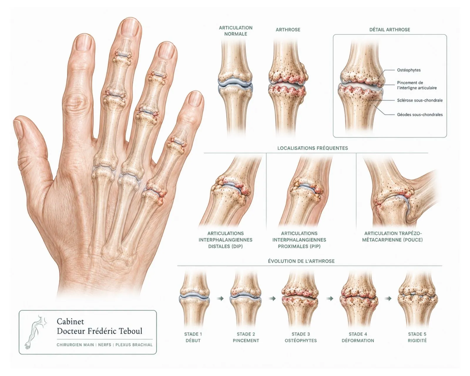
Each finger has three joints. Primary osteoarthritis of the hand has very consistent targets: it mainly affects the joint closest to the nail, somewhat less the middle joint, and largely spares the one that connects the finger to the palm.
This pattern is not a minor detail: it is one of the features that helps distinguish osteoarthritis from an inflammatory disease, which does not affect the same joints.
The thumb base is also affected, but its mechanics, progression and treatment are so distinctive that it deserves an article of its own. See the thumb-base osteoarthritis article.
Nodes: what these bumps really are
The hard lumps that appear on the top of the joints are neither deposits nor temporary swelling. They are bony outgrowths — the bone reacts to cartilage wear by remodeling along the edges of the joint.
Heberden's nodes
On the joint closest to the nail.
Bouchard's nodes
On the middle joint of the finger.
They are permanent: once formed, they do not go away. But their appearance says nothing about pain — some people have many and feel little, others the opposite.
"Is this rheumatoid arthritis?"
This is the first worry, and it deserves a clear answer rather than a reassuring phrase.
The European guidelines on the diagnosis of hand osteoarthritis established a key point: no single test is enough to make the diagnosis on its own. It is the combination of features that matters. EULAR 2009
Why your doctor asks about your mother's hands
Probability of hand osteoarthritis when Heberden's nodes are the only finding.
With three additional features: age over 40, a family history of nodes, and joint-space narrowing visible on an X-ray of any finger.
Hence the seemingly trivial questions about when it started and about your parents' hands: they make the diagnosis about four times more reliable.
A few markers help tell the two situations apart, without replacing an examination:
Morning stiffness is brief in osteoarthritis. Prolonged stiffness, lasting hours, points elsewhere.
The pain is mechanical: it increases with use and eases with rest.
The joints affected are not the same. Primary osteoarthritis usually spares the ones that connect the fingers to the palm.
Blood tests are normal in osteoarthritis. They are used precisely to rule out an inflammatory disease.
The erosive form: a variant worth knowing about
There is a less common, more inflammatory form of this osteoarthritis. It shows up as red, warm and markedly more painful joints, in flare-ups, with a significant impact on function. On X-ray, it produces distinctive images, different from ordinary osteoarthritis.
This is the form most often mistaken for an inflammatory disease — and it is also the one that most warrants active management rather than simple monitoring.
What finger osteoarthritis causes — beyond the pain
The functional difficulty involves fine movements: buttoning a garment, writing, turning a key, sewing, opening a package. These are not feats of strength, they are matters of precision — and that is what makes this osteoarthritis more disabling than it looks.
A rarely discussed aspect
Finger deformity also has a visible dimension. Many people suffer from it without daring to say so, because they feel it is not a legitimate medical concern.
Indeed, the hand is on constant social display: deformities, sometimes very pronounced, cause a cosmetic distress that outweighs the functional difficulty.
This is a legitimate concern. Distress about the appearance of the hands is something that should be heard during a consultation, just like pain — even if, on its own, it does not change whether treatment is indicated.
Treatments: what works, and what works little
This is where precision matters, because many things are offered and few have been shown to produce an effect the patient actually feels.
The systematic literature review that informed the 2018 European guidelines assessed each option against a threshold of clinically noticeable difference. The result is telling: only three treatments cross that threshold across hand osteoarthritis as a whole — prolonged wear of a splint at the thumb base, oral anti-inflammatories, and corticosteroid injections into the finger joints. EULAR review 2018
One fact to know before any decision: no treatment slows the progression seen on X-rays. They all act on symptoms. None repairs the cartilage or stops the process.
What comes first
The European guidelines put first education in ergonomic principles, pacing of activities and assistive devices, together with exercises to improve function, build muscle strength and reduce pain.
In practical terms: use the large joints rather than the small ones, add wider grips to utensils, get a jar opener. These are modest measures whose combined effect is real — provided they are kept up, because the benefit of exercises fades once they stop.
Medications
Topical treatments are preferred over systemic ones, and anti-inflammatory gels are the first choice.
Oral pain relievers, especially anti-inflammatories, should be considered for a limited time.
Chondroitin sulfate may be used to relieve pain and improve function. The systematic review notes, however, that this finding rests on a single trial.
Disease-modifying antirheumatic drugs should not be used.
The guidelines also advise against heat therapy, ultrasound and hyaluronic acid injections.
Injections: here, the opposite of the thumb base
This is the most interesting point in this article, and it shows why a guideline can never be boiled down to a slogan.
Two neighboring joints, two opposite conclusions
The European guidelines state that corticosteroid injections should generally not be used in hand osteoarthritis, but may be considered in patients with painful interphalangeal joints.
The reason for this distinction is explicit: the recommendation was fully revised because new data could not confirm an effect greater than placebo at the thumb base, whereas a trial on painful finger joints showed superior effectiveness.
In other words: what is not supported for the thumb is supported for the fingers. See the thumb-base osteoarthritis article.
The wording "should generally not" was chosen deliberately: the task force acknowledged that in certain situations — particularly when joint inflammation is obvious — injection remains a treatment option.
These injections into very small joints require a precise technique.
Surgery: when, and which type
The European guidelines are explicit: surgery should be considered for structural abnormalities when other treatments have not relieved the pain enough. For the finger joints, the options mentioned are fusion (arthrodesis) or arthroplasty.
These two words cover two opposing philosophies.
The choice depends on the finger involved, the alignment of the joint, the quality of the bone and, above all, on what you do with your hands. There is no single right answer. Each finger has a very specific function, and the treatment must be tailored to the joint involved and to the patient's functional demands.
An honest methodological caveat: the European systematic review notes that no trial has compared surgery with a sham procedure or with non-operative treatment. Surgical results therefore rest on case series, not on controlled trials.
After a procedure
After arthroplasty, the post-operative course depends on the technique used and the implant. For my part, I use an anterior approach that allows early rehabilitation. The procedure is done as an outpatient under regional anesthesia. Post-operative rehabilitation is as important as the surgery itself, and must last at least three months after the procedure.
After a distal interphalangeal fusion, there is no splint or immobilization. Rehabilitation of the finger begins immediately: the fusion is fixed with a cannulated titanium screw, an extremely solid construct.
What will happen over time
Finger osteoarthritis progresses slowly, over years. The nodes settle in permanently, but the pain does not follow the same course: it is often at its worst while the deformities are forming, then eases once the joint has stiffened.
This is an important point: a deformed hand is not necessarily a painful hand, and the passage of time is not only about things getting worse.
Frequently asked questions
Glossary
A bony outgrowth on the joint closest to the nail.
A bony outgrowth on the middle joint of the finger.
A bony formation that develops along the edge of a worn joint.
Permanent surgical fusion of a joint in a chosen position.
Replacement of a joint with an implant, in order to preserve mobility.
A more inflammatory form of finger osteoarthritis, with marked painful flare-ups.
References
Zhang W, Doherty M, Leeb BF, et al. EULAR evidence-based recommendations for the diagnosis of hand osteoarthritis: report of a task force of ESCISIT. Ann Rheum Dis. 2009. PMID 18250111 — 10 propositions, Delphi method, 15 European countries
Kloppenburg M, Kroon FPB, Blanco FJ, et al. 2018 update of the EULAR recommendations for the management of hand osteoarthritis. Ann Rheum Dis. 2019;78(1):16-24
Kroon FPB, Carmona L, Schoones JW, Kloppenburg M. Efficacy and safety of non-pharmacological, pharmacological and surgical treatment for hand osteoarthritis: a systematic literature review informing the 2018 update of the EULAR recommendations. PMC6203105
Randomized trial on the efficacy of triamcinolone hexacetonide injection in the interphalangeal joints, 12 weeks. J Rheumatol. 2015;42:1869-1877
Prevalence data used
Modified Kellgren-Lawrence score — stage boundaries
Outcomes of interphalangeal arthroplasties — ranges of motion and revision rates
Characterization of erosive osteoarthritis and radiographic criteria
Content produced, reviewed and approved by Dr Frédéric Teboul, surgeon of the hand, brachial plexus and peripheral nerves, member of the Académie Nationale de Chirurgie.
Last updated: August 20, 2026
Disclaimer. This article is intended for information only. It does not replace a medical consultation. Only a clinical examination can establish a diagnosis and choose a treatment suited to your situation.
Author of this publication
Every piece of content is signed by its author and reviewed before publication.
 Author
Dr Frédéric Teboul
Hand surgeon — specialist in the brachial plexus and peripheral nerves
Author
Dr Frédéric Teboul
Hand surgeon — specialist in the brachial plexus and peripheral nerves